Contents
Ultraviolet absorbance of oligonucleotides
Measuring the UV absorbance
The heterocyclic bases of DNA are aromatic and absorb in the ultraviolet region of the electromagnetic spectrum. The λmax for all Watson-Crick bases is between 250 nm and 280 nm. By measuring the absorbance (also referred to as optical density or OD) of a buffered aqueous solution of an oligonucleotide at 260 nm it is possible to estimate the concentration and quantity of the oligonucleotide present in solution.
The Beer-Lambert law states that the absorbance, A, of a sample, is equal to the product of the extinction coefficient, E, the concentration, c, and the length of the UV cell (or cuvette), l:
The absorbance of an oligonucleotide can be measured easily by placing a solution of the oligonucleotide (typically diluted) in a UV cell inside a UV spectrometer. This value (a dimensionless quantity) is often described as the "OD reading", and gives a rough-and-ready indication of the concentration of the sample.
In order to determine the concentration it is necessary to know the extinction coefficient. To get an accurate value, the extinction coefficient must be measured experimentally, but in practice it is possible to predict the extinction coefficient with good accuracy.
Extinction coefficients of oligonucleotides
There are two simple methods that are used commonly for the prediction of extinction coefficients: the base composition method and the nearest-neighbour method.
Base composition method
The base composition method simply sums the extinction coefficients for the isolated nucleosides (A, C, G and T) to give an approximation for the extinction coefficient of the oligonucleotide. This sum has to be multiplied by a factor of (typically) 0.9 to account for base stacking, which decreases the extinction coefficients of the bases within the oligonucleotide relative to the free nucleotides.
where is the number of bases of type present. For RNA, the formula is
For example, for a DNA oligonucleotide, dAGCGT, the extinction coefficient can be calculated as
Because the base composition model depends only on the isolated nucleosides, the calculation can easily accommodate modifications. If, in the above sequence, the A is replaced by 8-bromo-dA (E = 14.8), the calculation becomes
Nearest-neighbour method
The nearest-neighbour model [1] takes into account interactions between adjacent bases (and between the "ends", E, of the sequence). This can be done by simply summing over the interaction terms including ends (see below), or by summing over (different) interaction terms and subtracting terms for individual nucleotides. These nearest-neighbour parameters are measured empirically, and can give very accurate values for extinction coefficients of DNA. For the above sequence, the calculation is
A problem with this method is that modifications (whether internal or terminal) are not easily accommodated. For example, if the C in the above sequence is replaced by 5-Me-dC, the nearest-neighbour parameters GC and CG are unknown. Because the parameters are determined empirically, there is no direct link between the extinction coefficient of the isolated nucleoside and its nearest-neighbour parameters. The problem is exacerbated when multiple modifications are present.
Comparison of methods
For unmodified oligonucleotides, the agreement between the base composition and nearest-neighbour models is surprisingly good for most oligonucleotides. In general, the nearest-neighbour method is preferred for unmodified DNA, and can give very accurate extinction coefficients. The base composition model is a good substitute, and is invaluable when dealing with modified DNA.
The two methods do diverge, however, for certain types of sequences (for example, sequences with long stretches of A bases). The methods can be compared easily using the oligo calculator.
Hypochromicity and double-stranded DNA
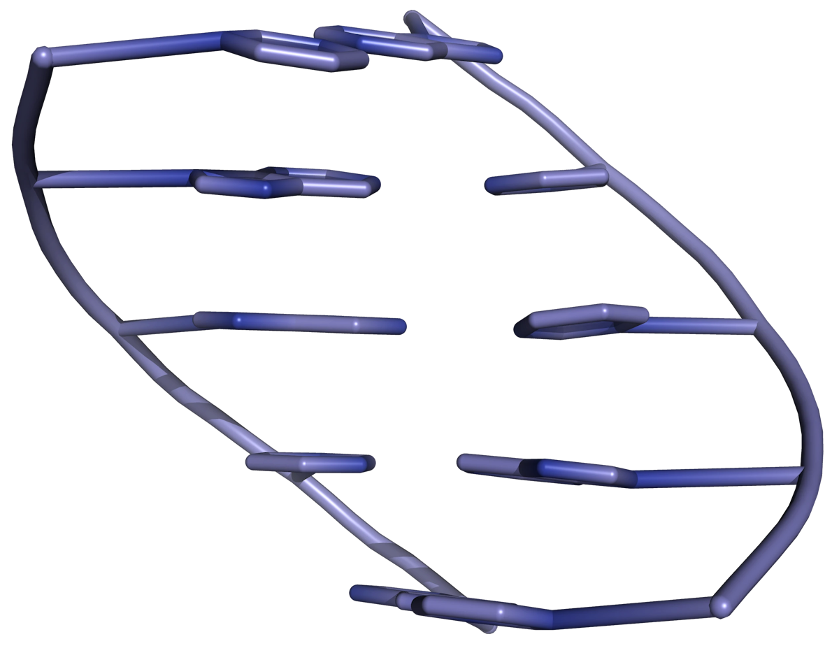
When calculating the extinction coefficient of double-stranded DNA (composed of an oligonucleotide and its reverse complement), the extinction coefficients of the two constituent strands (i.e. the sequence and its complement) are used in the calculation. The absorbance of a DNA duplex is always lower than the sum of the absorbances of the two individual strands, owing to π-stacking of bases in the double helix (Figure 1). This reduction in absorbance is called hypochromicity.

This hypochromicity must be quantified when calculating the extinction coefficient of double-stranded DNA. The hypochromicity h can be estimated by the simple formula
where fGC is the fraction of G and C bases in the sequence, and fAT is the fraction of A and T bases. This results in values of h around 0.2. The extinction coefficient of double-stranded DNA must be reduced by this amount in the calculation:
For example, for the DNA duplex composed of the oligonucleotide ATTTGCG and its reverse complement CGCAAAT, the extinction coefficient is calculated as
Other wavelengths
Wavelength of maximum absorbance
So far the discussion has concentrated on extinction coefficients calculated at 260 nm. This wavelength is used because the UV spectrum of a typical oligonucleotide reaches a maximum at around this wavelength. The maximum absorbance λmax can be estimated (for DNA oligonucleotides) using the formula
This formula gives values of λmax around 260 nm unless the sequence has an unusually high G content.
UV Spectrum
Extinction coefficients at wavelengths other than 260 nm can be calculated from those measured or estimated at 260 nm by using empirically derived nearest-neighbour parameters. Using this method, an entire UV spectrum can be predicted, although that is beyond the scope of this article.
Calculating concentration, mass, yield
Once the extinction coefficient has been determined, the concentration of an oligonucleotide can be calculated by dividing the absorbance (OD reading), A, by the extinction coefficient, E. Assuming that the length of the UV cell is 1 cm, Eqn. (1) gives
The total amount of DNA can then be calculated by multiplying the concentration by the volume of the cell. This allows the percentage yield to be determined.
Example
An oligonucleotide with the sequence dGGACACGAGGTCTT was synthesized on the 1.0 μmol scale. After synthesis and purification, a 1.5 mL solution of the oligo in water was obtained. A 50 μL aliquot of the solution was taken and made up to 3 mL of water. The absorbance of the solution was measured at 0.762 OD units (at a wavelength of 260 nm).
Calculation of extinction coefficient
In order to calculate the concentration from the OD reading, we need to know the extinction coefficient. We can estimate it using the base composition model formula (Eqn. (2)):
Calculation of concentration
When we took the OD reading, we took a 50 μl aliquot and made the volume up to 3 mL, so we must multiply the OD reading by a factor of 3000/50:
We can now calculate the concentration of the oligo by dividing the measured absorbance (A260) value by the calculated extinction coefficient (E260):
Calculation of amount
We can calculate the amount of DNA by multiplying the concentration by the volume of DNA obtained:
Calculation of yield
We have calculated the amount of oligo synthesized, and we know that the synthesis scale was 1 μmol. The ratio of these values gives the percentage yield:
This might not look great, but it's not bad for 20 detritylation reactions, 19 coupling reactions, 19 capping steps 19 oxidation steps an ammonia cleavage step and the simultaneous deprotection of 19 phosphates and 15 bases!
UV melting
UV (ultraviolet) melting is the most common method for determining DNA duplex stability. On transition from double-stranded to single-stranded DNA, there is a significant increase in UV absorbance. This is a direct result of hypochromicity, a lowering of UV absorbance in the double helix as a result of base stacking.
The DNA duplex is dissolved in an aqueous buffer and heated slowly until it dissociates (or "melts") into two single strands. Heating double stranded nucleic acids causes the strands to unwind (denature) by disrupting the ordered stacking of the bases and breaking hydrogen bonds. A graph of UV absorbance (A) against temperature (T) gives a characteristic sigmoidal curve (Figure 2).
The mid-point of the melting curve is called the melting temperature, or Tm. The Tm gives an indication of duplex stability, and can be used to determine thermodynamic parameters for melting. This UV melting data can provide valuable information about the stability of mismatched base pairs which can result from mutagenesis.
References
[1] Predicting ultraviolet spectrum of single stranded and double stranded deoxyribonucleic acids. Biophys Chem 2008, 133, 66.